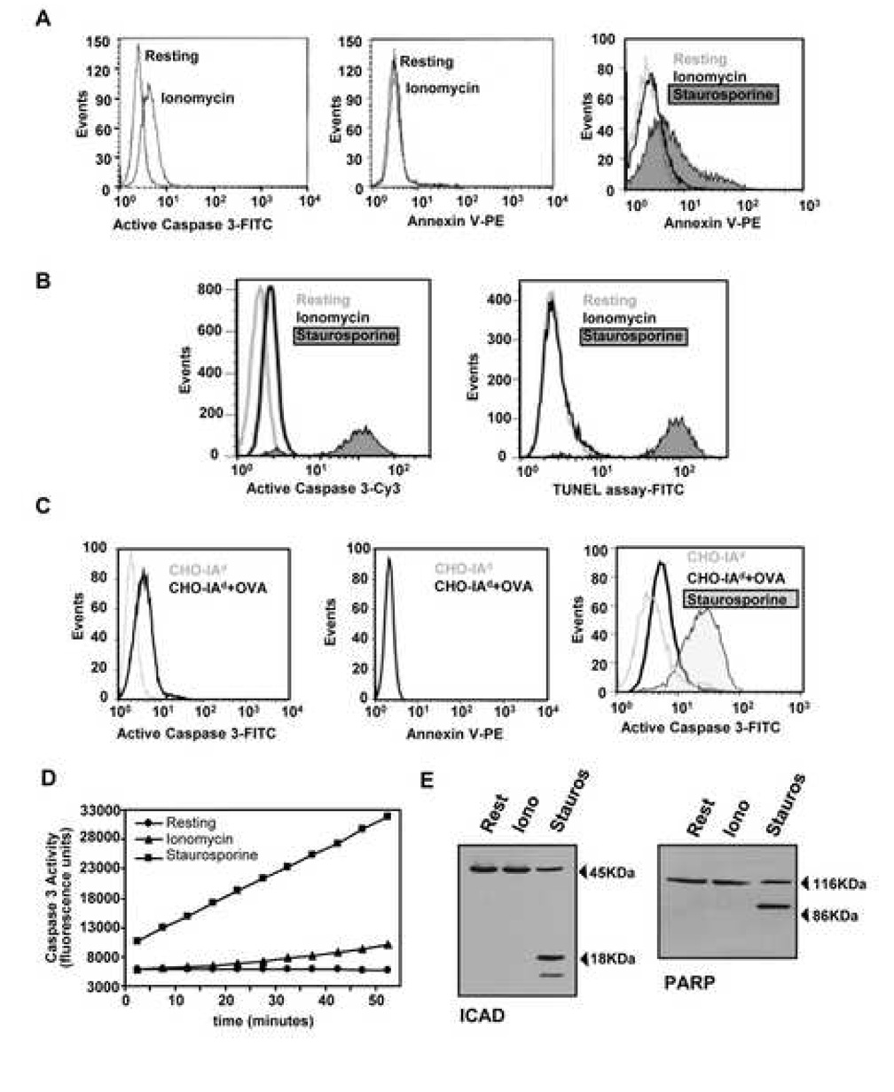
Figure 2. Activation of Caspase 3 does not induce apoptosis in anergic T cells.
A. DO11.10 Th1 cells treated with ionomycin or left resting for 12 hours. T cells were then recovered, washed, stained with an antibody against the active form of caspase 3 (left panel) or with Annexin V-PE (center panel) and analyzed by FACS. T cells treated with staurosporine (1µM, 4 hours) were used as positive control for Annexin V-PE staining (right panel). B. Levels of apoptosis were also detected by TUNEL assay in Th1 cells treated with 1µM ionomycin for 12 hours or with staurosporine for 4 hours. The same cells were also stained for active caspase 3 (left panel). C. Similar analyses were performed in T cells cultured for 12 hours with CHO-IAd cells loaded or not with OVA. D. Caspase 3 activity was measured using a fluorogenic caspase-3 substrate in cell lysates from Th1 cells left resting, anergized with ionomycin for 12 hours or treated with 1µM staurosporine for 4 hours. E. Th1 cells were left resting, incubated with 1µM ionomycin for 12 hours or treated with 1µM staurosporine for 4 hours. Total cell lysates were and analyzed by western blot using antibodies that recognized the proapoptotic caspase 3 substrates PARP and ICAD.

