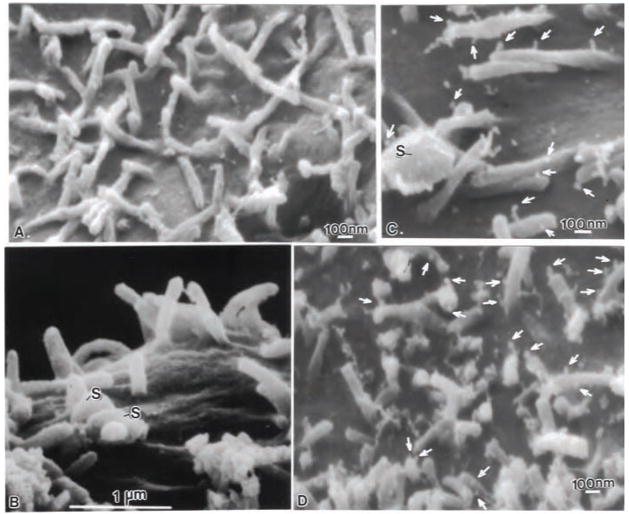
Fig. 4.
FESEM images of Caco-2 cell microvilli incubated for 2.5 hr with mAb-nanoloops in the presence and absence of B. cereus spores. (A) Following incubation with mAb -74.5.2 functionalized nanoloops, the microvillous surface showed no marker uptake in the absence of spores. (B) Caco-2 microvilli incubated with B. cereus spores (s) and control IgG1k-nanoloops showed no marker uptake, even on microvilli adjacent to attached endospores. (C) However, numerous mAb-nanoloops (arrows), attached to microvilli and the exosporia of B. cereus spores. (D) At lower magnification, the extensive localization of gC1qR/p33 with mAb -nanoloops (arrows) is apparent on the microvilli adjacent to B. cereus spores.

