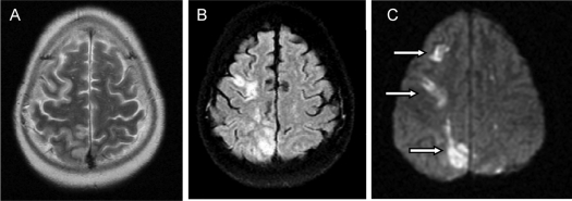
Figure 2.
Brain images obtained 2 d after surgery using identical techniques as in Fig. 1. A, T2-weighted images show no definite brain lesions. However, FLAIR (B) as well as diffusion-weighted (C) images reveal abnormal areas of increased signal intensity in the right cerebral hemisphere (arrows). Corresponding low-signal-intensity lesions were present on the apparent diffusion coefficient maps (not shown). The combination of these signal changes indicates the presence of acute ischemic infarctions.

