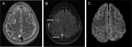
Figure 3.
Brain MRI obtained 2 yr after surgery. A and B, T2-weighted (A) and FLAIR (B) images show small glial scars in two of the ischemic infarctions previously identified in the right cerebral hemisphere; the third infarction had completely resolved. C, Diffusion weighted image at the same level is normal indicating good healing and no evidence of new acute lesions.

