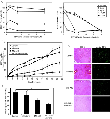
Figure 6.
IGF-1R antagonists enhance the inhibitory effects of mitotane. A, RL251 (left panel) and H295 (right panel) cells were incubated in triplicate with a combination of mitotane and increasing concentrations of NVP-AEW541. Proliferation was assessed with MTS reagent. Data are representative of three independent experiments and displayed as mean ± sd. B, Mice harboring H295 xenografts were randomized into four groups (n = 20 per treatment arm) and treated with vehicle or IMC-A12 every other day and/or mitotane once daily for the duration of the experiment. Tumor volumes were measured three times a week. Data are presented as log ratios of tumor size over initial tumor size means ± se. C, Hematoxylin and eosin (H & E)-stained section of H295 tumor xenografts (left panels) at ×40 magnification. Lectin-FITC immunohistochemical analysis (right panels) was performed to detect relative levels of endothelial cells. D, Quantitative RT-PCR of three or four tumors from each treatment arm with primers detecting all four isoforms of human VEGF. The y-axis represents relative VEGF levels normalized to glyceraldehyde 3-phosphate dehydrogenase transcript levels followed by normalization to control tissue values. Each value represents the average of triplicates of two independent experiments and data are presented as the mean ± se. *, P < 0.05.

