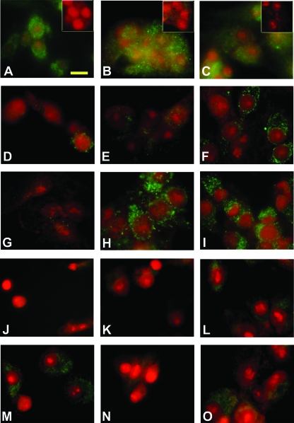
Figure 5.
Immunofluorescent detection of EP1, EP2, EP3, tPA, and PAI-1 proteins in cultured granulosa cells. For EP protein detection, granulosa cells obtained from animals undergoing controlled ovarian stimulation before hCG administration were maintained in vitro with hCG plus indomethacin for 36 h. The cells were formalin fixed and stained (bright green) for EP1 (A), EP2 (B), and EP3 (C) proteins. Immunofluorescence was reduced or eliminated when primary antibodies were preincubated with the respective blocking peptide (A–C, insets). For immunofluorescent detection of tPA and PAI-1 proteins, granulosa cells were cultured as described in Fig. 4A with no treatment (D and J) or with hCG plus indomethacin (E–I and K–O). After 24 h, some cultures received additional treatment with PGE2 (F and L), 17 phenyl-trinor PGE2 (G and M), butaprost (H and N), or sulprostone (I and O). Cells were cultured for a total of 28 h for immunodetection of tPA (D–I) or 36 h for immunodetection of PAI-1 (J–O). Immunodetection appears green; nuclei are counterstained red. Images in A–C are representative of two animals. Images in D–O are representative of three to four animals per treatment. Scale bar in A, 20 μm (all images).

