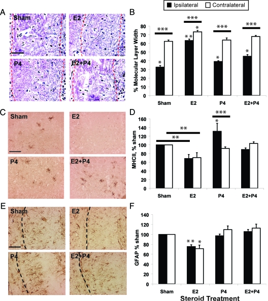
Figure 2.
P4 attenuates E2-induced compensatory sprouting after ipsilateral ECL (see Fig. 1). All rats were ovariectomized before ECL and killed 14 d after ECL. Controls received sham pellets without steroids. Scale bars, 100 μm. n = 8 rats per group, 3 sections per brain. Results are means ± sem. A, Axonal fibers (Holmes stained) in the ipsilateral molecular layer of the dentate gyrus (borders outlined by red dashed lines). B, Average fiber band width in molecular layers of the ipsilateral (lesioned) and contralateral (unlesioned) sides, expressed as percent total molecular layer width (granule neuron layer edge to fissure). *, P < 0.05 and **, P < 0.001 from other groups in respective hippocampal side; ***, P < 0.0001, ipsi- vs. contralateral side. C, Microglial activation in the hippocampal hilus by OX6 immunostaining (epitope for MhcII 1a peptides). D, OX6-immunostained area in ipsi- and contralateral hilus, as percentage of sham. *, P < 0.05 and **, P < 0.001 from other treatments on respective side; *** P < 0.01, ipsi- vs. contralateral side. E, Astrocyte GFAP immunostaining in the ipsilateral molecular layer of the dentate gyrus. Dashed line shows the hippocampal fissure. F, GFAP-immunostained area in ipsi- and contralateral hippocampus, as percentage of sham. *, P < 0.01 and **, P < 0.005 vs. other treatments on respective hippocampal side.

