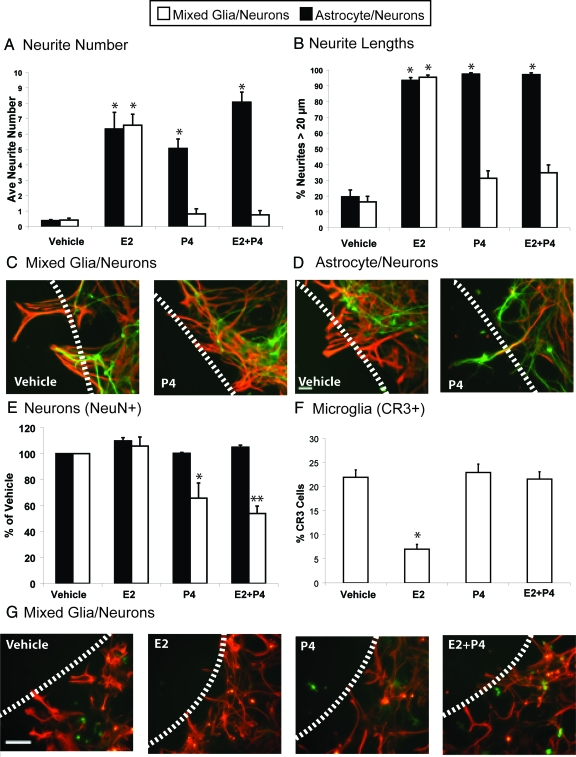
Figure 3.
Wounding-in-a-dish model of lesions to evaluate whether the presence of microglia influences responses of neurite outgrowth in the presence of E2 and P4 in culture media. Glia were cocultured with E18 neurons for 3 d and subjected to scratch wounding, with subsequent analysis 48 h later. Green, neurite MAP-5 immunostaining; red, astrocytic GFAP immunostaining; scale bar, 100 μm; dashed line, scratch wound zone. Results show the average of three experiments ± sem. A, Numbers of neurites extended into the wound zone per 0.5 mm2 in cultures of astrocytes (black bars) or mixed glia (white bars). *, P < 0.001. B, Neurite outgrowth, expressed as percentage of neurites bigger than 20 μm (legend as for A). *, P < 0.001. C, Addition of P4 alone did not induce neurite outgrowth in mixed glia-neuron cocultures containing 30% microglia and 70% astrocytes. D, Addition of P4 alone induced neurite outgrowth in astrocyte-neuron cocultures. E, NeuN-positive neurons in 0.250 mm2 adjacent to wound zone. *, P < 0.0005, vehicle and E2 in respective coculture model; **, P < 0.0001, vehicle and E2. F, Histogram of microglia adjacent to wound zone showing percentage of cells immunoreactive for CR3/total glial cells (CR3+GFAP); *, P < 0.0001 from other treatments. G, Glia in mixed glial-neuron cocultures with steroid treatment. Immunostaining for astrocytes GFAP (red) and microglia CR3 (OX42 antibody, green).

