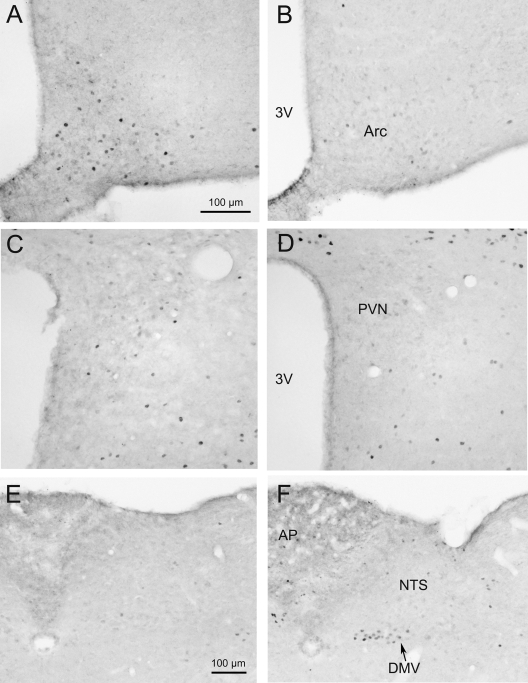
Figure 5.
Microphotographs of Fos immunoreactivity in the Arc, PVN, and dorsal-vagal complex (DMV and NTS) of overnight fasted CRF-OE (right panels) and WT (left panels) mice. Overnight fasted CRF-OE mice showed much less Fos-ir neurons in the Arc (B) compared with WT mice (A). No difference was detectable in the PVN (C and D) and NTS (E and F). Overnight fasting resulted in a significantly increased number of Fos-ir neurons in the DMV of CRF-OE mice (F) compared with WT mice (E). The scale bar in photo A represents the magnification for photos A–D and the one in E for E and F. 3V, Third cerebral ventricle; AP, area postrema.

