
Figure 1.
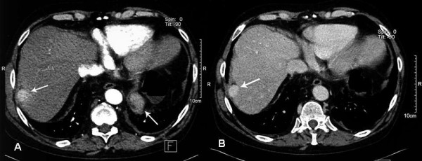
CT scan. (A) Axial IV contrast enhanced CT. Arterial phase image showing a 2 × 2.7 cm hypervascular, subcapsular nodule in segment VII of the liver. (B) Portal venous phase image.
Official websites use .gov
A
.gov website belongs to an official
government organization in the United States.
Secure .gov websites use HTTPS
A lock (
) or https:// means you've safely
connected to the .gov website. Share sensitive
information only on official, secure websites.
CT scan. (A) Axial IV contrast enhanced CT. Arterial phase image showing a 2 × 2.7 cm hypervascular, subcapsular nodule in segment VII of the liver. (B) Portal venous phase image.