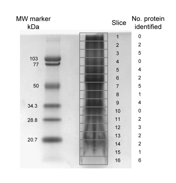
Figure 4.

1-D SDS-PAGE gel of developing white lupin root microsomal proteins. One hundred μg microsomal proteins were solubilized in SDS-PAGE sample buffer and separated on a 4–15% gradient polyacrylamide gel. The protein gel was stained with Coomassie blue R-250. Sixteen 5 mm slices were excised from the 1-D gel as outlined. The gel slices were digested with trypsin and subjected to LC-MS/MS analysis.
