Figure 2.
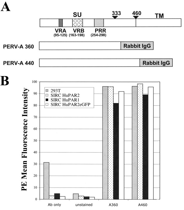
In vitro PERV SU-IgG binding by HuPAR1 and HuPAR2. (A) shows the SU constructs of either minimally-required (360 a.a.) or full-length (440 a.a.) PERV-A envelopes. All SU-IgG constructs contain Variable Region A (VRA), Variable Region B (VRB) and the Proline Rich Region (PRR). Binding of the soluble SU-IgG constructed is detected by a PE-conjugated secondary antibody that recognizes Rabbit IgG. (B) shows the Mean Fluorescence Intensity (MFI) detected by FACS and is representative of duplicate experiments. The 293 T cell line (gray bars) is a positive control for PERV-A binding. SIRC HuPAR2 (dotted bars) is a control for interference of the eGFP epitope tag in PERV-A binding. Both HuPAR1eGFP (black bars) and HuPAR2eGFP (white bars) bind PERV-A 360 and PERV-A 440 similar to the levels of 293 T and SIRC HuPAR2. Therefore, the difference between HuPAR1 and HuPAR2 in PERV-A 14/220* infection is not due to any difference in envelope binding.

