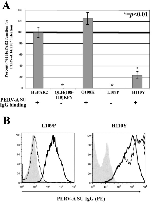
Figure 5.
Contribution of QLH(108–110) to HuPAR2 function for PERV-A 14/220* infection at the single residue level. The individual requirement of each residue of the QLH(108–110) region to PERV-A binding and infection was determined. (A) shows both the percent (%) of wild-type (WT) HuPAR2 function and full-length PERV-A SU binding for QLH(108–110)KPY, Q108K, L109P and H110Y. The L109P mutant does not bind PERV-A SU. H110Y results in a significant decrease (p < 0.01) of HuPAR2 function for infection, but does not affect PERV-A SU binding. (B) shows the FACS histogram from the binding assay for both L109P and H110Y. The negative controls (naïve SIRC cells; gray shading) and L109P (dotted black line) shown in the first plot, indicate no binding of PERV-A SU IgG compared to HuPAR2eGFP (solid black line). In the second plot, H110Y (dotted black line) and the positive control, HuPAR2eGFP (solid black line) show equivalent SU IgG binding. Therefore, L109 is the only residue within the QLH mini-region that determines HuPAR2 binding.

