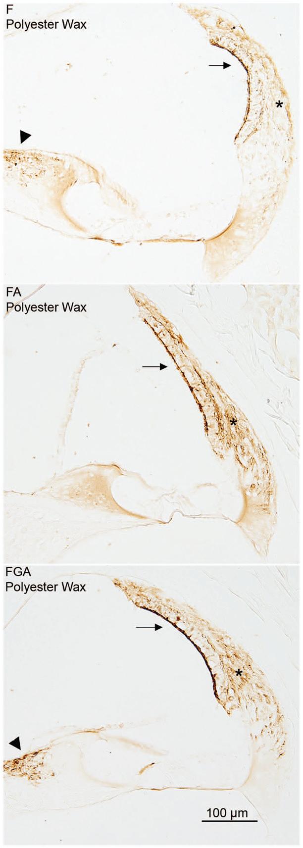
Fig. 2. Immunostaining for PGDS.

The effect of fixative is demonstrated in this panel. The PGDS antibody stains the fibrocytes of the spiral limbus (arrowhead), the type I fibrocytes (asterisk) of the spiral ligament and the marginal cells of the stria vascularis (arrow). In all three images shown in the panel, the embedding medium was polyester wax. The immunostaining improves as one progresses from the F fixed tissue to the FA tissue to the FGA tissue.
