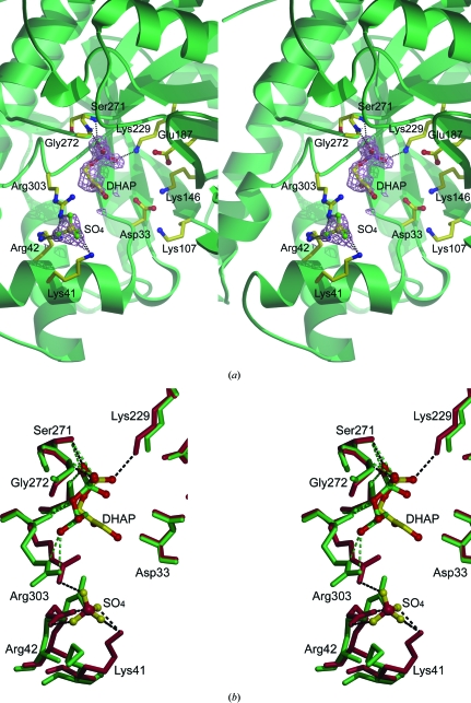
Figure 6.
Stereoview of the active site of fructose 1,6-bisphosphate. (a) DHAP and a sulfate ion depicted in ball-and-stick representation along with conserved active-site residues in D128V aldolase. A 1σ electron-density map is shown (violet cages). (b) Superposition of the active-site residues of D128V aldolase in red with DHAP and sulfate ion in yellow and the residues of the wild-type DHAP-bound structure (Blom & Sygusch, 1997 ▶; PDB code 1ado) in green. Hydrogen bonds are shown in black for D128V aldolase and in green for the wild-type structure.

