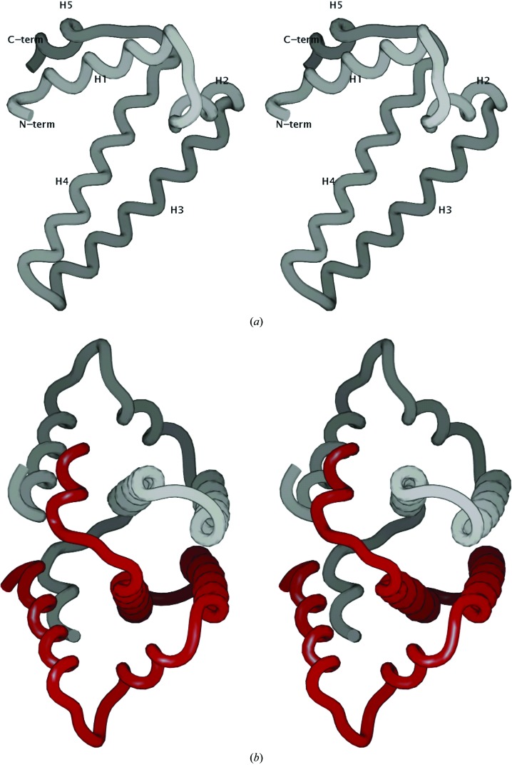
Figure 2.
(a) Backbone representation of the fold of Mtb HisE (residues 7–93). Helices H1–H5 are labeled. All molecular graphics in this paper were produced using SPOCK (Christopher, 1998 ▶). (b) Backbone representation of the dimer formed by two subunits packing together to form a four-helix bundle. Helix H5 in the C-terminus of each subunit wraps around the other subunit and contacts helix H1, interlocking the dimer.

