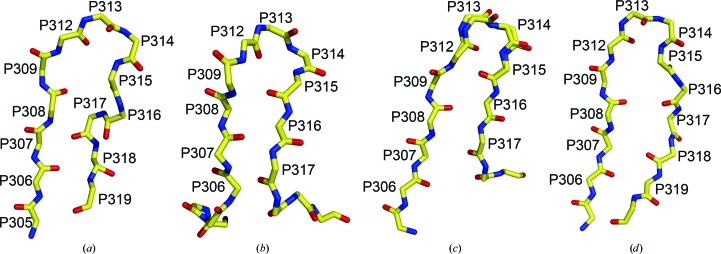
Figure 6.
Comparison of V3 crystal structures. The Cα-backbone structures are shown in a similar orientations for (a) the UG1033 V3 peptide bound to Fab 447-52D (b) the UG1033 V3 peptide bound to Fab 2219 (PDB code 2b1a) and the V3 loops from two gp120 core complexes: (c) PDB code 2b4c and (d) PDB code 2qad. Two conformations were observed for the V3 crown in the core gp120 V3 structure with PDB code 2b4c and both of these conformations are shown in (c).

