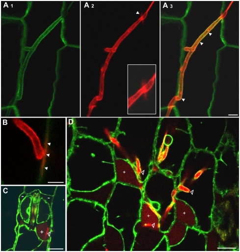
Figure 5. Secretion of Pep1-mCherry into the maize apoplast.
A, B: SG200pep1M growing intracellularly in epidermal cells of maize line ZmPIN1a-YFP, 48 hpi. A1, A2 and A3 show the same hyphae with PIN1-YFP (green), Pep1-mCherry (red) and the merged yellow signals indicating co-localisation (arrowheads) around fungal hyphae, respectively. At sites of cell-to-cell passages, Pep1-mCherry is spreading from the fungal hyphae (A2, insert; B). Bars: 5 µm. C, D: SG200pep1M growing intracellularly in epidermal cells of maize line ZmTIP-YFP, 48 hpi. Plasmolysis was induced by 1 M NaCl, collapse of vacuoles results in enlarged apoplastic spaces. In cells colonized by SG200pep1M, this space is filled by Pep1-mCherry (+) which is not the case in cells not colonized by the fungus (−). Bars: 15 µm.

