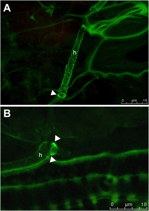
Figure 6. Immunolocalization of Pep1-HA in U. maydis infected maize tissue.
A, B: Confocal projections showing immunolocalization of Pep1-HA in maize tissue infected by SG200Δpep1-pep1HA. Pep1-HA is detected around intracellular hyphae (h), predominantly accumulating at sites of cell to cell passage (arrowheads). Bars are given.

