Figure 5.
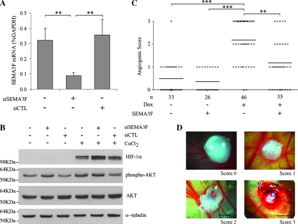
Physiological effects of SEMA3F loss. (A, B) H661 cells were transfected with siRNA against SEMA3F (siSEMA3F) or nontargeting siRNA (siCTL). Control was mock-transfected cells. (A) SEMA3F mRNA was monitored by quantitative real-time RT-PCR 48 hours after transfection. Values for three independent experiments, done in duplicate, are expressed in percentage of GAPDH expression. Bars, SD. (B) H661 cells were transfected by siRNA in absence or presence of 100 µM CoCl2 during 2 hours, and effects on signaling were monitored by Western blot analysis for HIF-1α, phospho-Ser473-AKT, and total AKT. α-Tubulin was used as a loading control. (C) Chorioallantoic membrane assays were performed to test angiogenesis. H358 FlpIn ZEB-1 cells were treated or not with Dox and then loaded on chick embryos in Matrigel with either presence or absence of recombinant SEMA3F-Fc. Six days after implantation, angiogenesis was evaluated independently by two observers with scores ranging from 0 to 3 as shown in D (scale, 3 mm). Using a nonparametric Kruskal-Wallis test, scores were significantly different with ZEB-1-induced cells versus noninduced cells (***P < .001) and ZEB-1-induced cells versus ZEB-1-induced cells plus SEMA3F (**P µ .01). Bars, mean. n, number of eggs.

