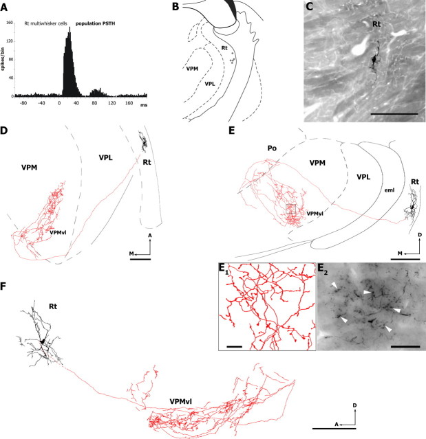
Figure 4.
Axonal projections of multivibrissa RT cells. A, Population PSTH of vibrissal responses recorded in six multiwhisker units (sum of all responses to all directions). Dots in B indicate the location of five multiwhisker RT units labeled with Neurobiotin. The RT cell labeled in C was reconstructed from serial sections, and drawings in D–F show horizontal (D), coronal (E), and sagittal (F) views of the dendritic and axonal arbors. Note that the axonal arbor is mostly restricted to the VPMvl, with a few collaterals in Po. E1, Axonal branches and boutons (arrowheads) within the framed area in E. Photomicrograph in E2 show a cluster of corticothalamic boutons in the VPMvl (arrowheads). A, Anterior; D, dorsal; eml, external medullary lamina; M, medial. Scale bars: B, 1 mm; C–F, 200 μm; E1, 10 μm; E2, 25 μm.

