
Figure 5.
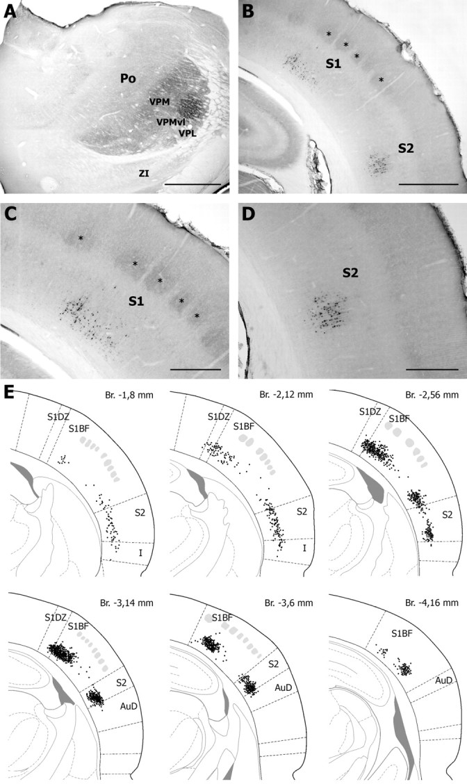
Retrograde labeling in the cerebral cortex after FluoroGold injection in the VPMvl. The injection site shown in A led to retrograde labeling of layer 6 cells in S1 and S2 (B). Clusters of labeled cells in S1 and S2 are shown at higher magnification in C and D, respectively. Asterisks indicate coreactive barrels. E, Distribution of retrogradely labeled cells in S1 and S2 on representative sections taken at different frontal planes (drawings adapted from Paxinos and Watson, 1998) [distance from the bregma (Br) as indicated; gray patches, barrels]. AuD; Dorsal auditory cortex; I, insular cortex; S1BF, barrel field of S1; S1DZ, dysgranular zone of S1; ZI, zona incerta. Scale bars: A, B, 1 mm; C, D, 500 μm.