Fig. 1.
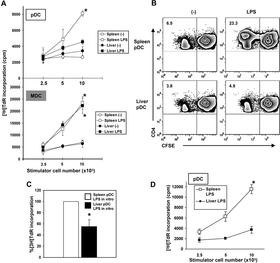
Liver pDC (immunobead-purified; mPDCA-1+) from LPS-injected C57BL10 (B10) mice induce significantly lower proliferation of allogeneic CD4+ T cells in 72h MLR, as determined by (A), [3H]TdR incorporation, and (B), CFSE dilution analysis compared with spleen pDC (*p<0.01). The numbers in the upper left quadrants indicate percent positive CD4+ T cells. A minimum of 20,000 CD4+ gated cells were analyzed. In comparison, LPS-stimulated mDC from spleen or liver induced much higher T cell proliferation compared with untreated liver mDC (A; *p<0.01). (C), pDC from liver or spleen were cultured overnight (16h) with LPS (1µg/ml), then used as stimulators in MLR. Results are expressed as relative [3H]TdR incorporation induced by spleen pDC as stimulators (*p<0.01). (D), Allostimulatory capacity of liver and spleen pDC from LPS-injected normal (non-Flt3L-mobilized) B10 mice (*, p<0.01). Data are representative of 5 (A and B) or 2 independent experiments (D), or means ± 1SD of 3 experiments (C).

