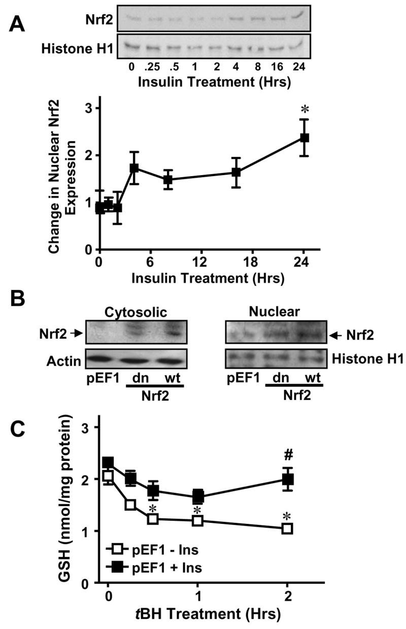
Figure 5. Cytosolic-nuclear distribution of Nrf2 and insulin stimulation of Nrf2 nuclear translocation and GSH recovery post tBH challenge.

A. Confluent IHECs were treated with 100 nM insulin for 24 hrs and nuclear protein was isolated at the indicated time points for assessment of nuclear Nrf2 expression by Western blotting (Top panel). Nrf2 expression was normalized to Histone H1 and expressed as fold change vs 0 hrs (Lower panel). * p<0.05 vs 0 hrs. B. Cytosolic and nuclear fractions were prepared from IHECs stably transfected with empty mammalian expression vector (vector control, pEF1), or with vector containing either dominant negative (dn) or wild-type (wt) Nrf2. Nrf2 protein contents in the subcellular compartments for each IHEC clones were examined by Western analyses. The membranes were reprobed with actin or histone H1, respectively to verify equal protein loading in the cytosolic and nuclear compartments. One representative of two Western blots is shown. C. IHEC cells stably transfected with an empty expression vector (pEF1) were treated with 100 nM insulin for 48 hrs prior to challenge with 100 μM tBH for 2 hrs. Cellular GSH contents were measured at designated times. * p<0.05 vs pEF1 minus Ins at 0 hrs. # p<0.05 vs pEF 1 minus Ins at respective time point.
