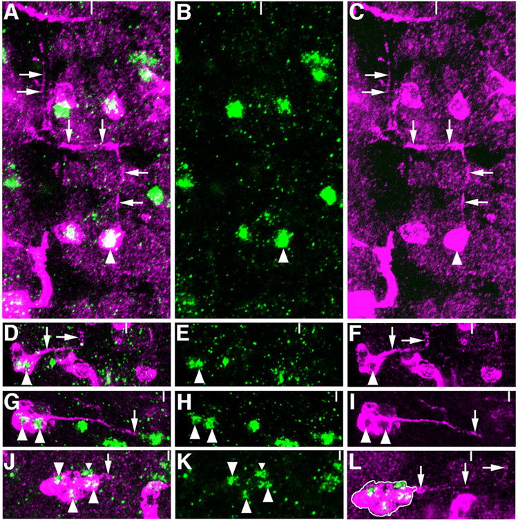
Figure 2.
Axon morphologies of clones including VG+ cells. A–C, One-cell GFP clone (magenta) in T3 includes the VG+ cell (green) of cluster iii (arrowhead). This cell projects an axon to the anterior in the ipsilateral connective in a stage 15 embryo. This axon turns at the anterior commissure, crosses the midline, and again projects anteriorally in the contralateral connective (arrows). D–F, GFP+ clone in A5 of a stage 17 embryo contains VG+ cells of cluster iv. This clone contains 9 cells, 4 of which are VG+. Two axons are extended by cells in this clone. An axon extended by a VG+ cell (arrowhead) reaches the outer edge of the longitudinal connective and turns to the anterior (arrows) just anterior to the level of the posterior connective. G–I, Second example of a clone including VG+ cells of cluster iv. This 8-cell GFP+ clone in T3 contains 5 VG+ cells. A single thick axon extended by two VG+ cells (arrowheads) reaches the midline at the level of the posterior commissure (arrows). J–L, Eight-cell GFP+ clone in A4 includes 3 VG+ cells of cluster v. An axon extended by this clone of cells crosses the midline and projects anteriorally in the contralateral connective (arrowheads). L is a photomontage to show the relationship of the clone shown in J with its axonal trajectory. One VG+ cell in cluster v is not included in this clone (J, small arrowhead).

