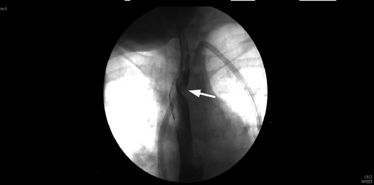
Figure 2.
Anteroposterior fluoroscopic image of esophagus demonstrating an oblique extrinsic defect coursing from inferior (patient’s left) to superior (patient’s right), consistent with an aberrant right subclavian artery at the upper thoracic level, just above the aortic arch. Arrow indicates filling defect

