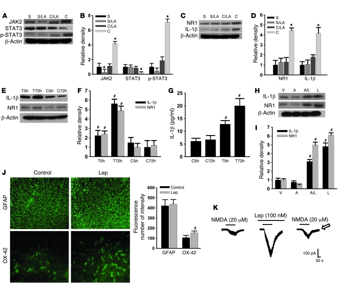
Figure 4. Role of JAK2 and STAT3 in the spinal leptin effect.
(A–D) The expression (Western blot) of JAK2, p-STAT3, NR1, and IL-1β was increased, whereas STAT3 was decreased on day 7 in CCI rats. The altered expression of JAK2, STAT3, p-STAT3, NR1, and IL-1β was prevented by once daily intrathecal administration of leptin antagonist (LA, 3 μg) for 7 days. S, sham; C, CCI; LA, leptin antagonist. (E and F) Exposure to leptin (100 ng/ml) for 5 or 72 hours upregulated NR1 and IL-1β expression in an organotypic spinal tissue culture (Western blot) and increased IL-1β in the culture medium (ELISA) (G). T5h, leptin exposure for 5 hours; T72h, leptin exposure for 72 hours; C5h, vehicle exposure for 5 hours; C72h, vehicle exposure for 72 hours. (H and I) Adding AG 490 (6 ng/ml) into leptin (100 ng/ml) culture medium for 72 hours significantly reduced leptin-dependent NR1 and IL-1β upregulation. V, vehicle; A, AG 490; L, leptin. (J) Exposure to leptin increased the expression of microglia (OX-42) in the spinal tissue culture. Scale bars: 120 μm (GFAP); 60 μm (OX-42). Histogram shows density measurement of immunofluorescent images (n = 5). (K) NMDA-induced current was enhanced in the presence of leptin. Arrow indicates recovery of NMDA current at 30 minutes after leptin washout. Data are shown as mean ± SD. *P < 0.05 versus sham; #P < 0.05 versus control.

