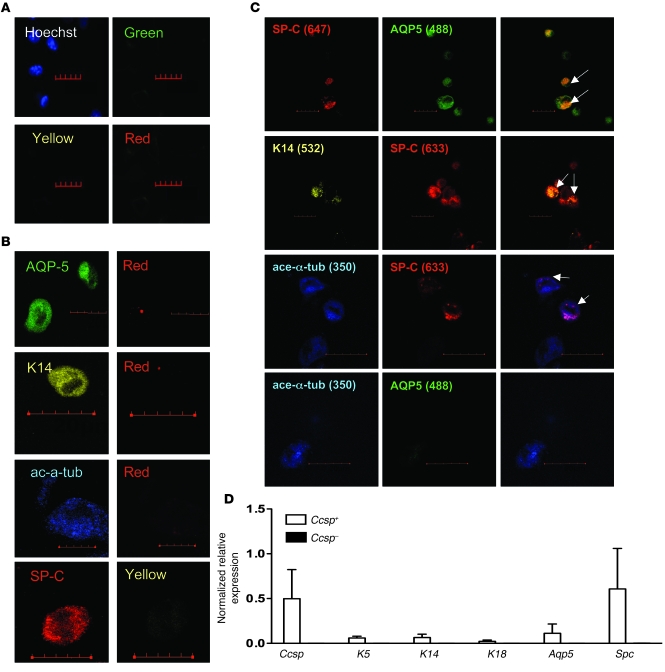
Figure 3. Epithelial multilineage differentiation of Ccsp+ BMCs.
(A) Isotype mix control staining of airway epithelial cells showed no cross-reactivity of the antibodies. (B) Control staining of isolated airway epithelial cells for each antibody. Bleed-through of signal is visualized in depicted representative images. Scale bars: 20 μm. (C) After 4 weeks in ALI culture, Ccsp+ cells gave rise to a multitude of epithelial lineage cells including proSpc+, proSpc+Aqp5+, proSpc+K14+, K14+, acetylated α-tubulin+, and acetylated α-tubulin+proSpc+ cells, whereas Ccsp– cells only gave rise to proSpc+ cells. White arrows point to double-positive cells. Hoechst counterstain was used to visualize nuclei (blue). Numbers in parentheses indicate the fluorescent emission wavelengths for the stains. Scale bars: 10 μm. Original magnification, ×40. (D) Real-time PCR confirmed expression of various epithelial genes in ALI-cultured Ccsp+ cells. Ccsp+ cells expressed various epithelial genes including Ccsp, K5, K14, K18, Aqp5, and proSpc, whereas Ccsp– cells only expressed proSpc. Gapdh was used as housekeeping gene for normalization of expression levels. Ccsp-sorted cells were cultured in a mixture of bone marrow– and epithelial cell–specific media and in ALI conditions to induce epithelial differentiation. Each bar represents normalized relative levels compared with tracheal epithelial cells. n = 4 sets of cells from 4 different animals.

