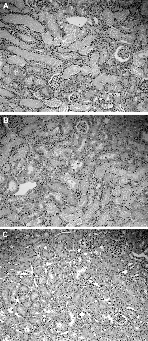
Figure 1.
Renal histological injury, as assessed at 3 days after unilateral ischemic injury. As a frame of reference for interpreting the biochemical data, 4-μm kidney sections were cut from 10% formalin-fixed tissues and stained with H&E. Extensive proximal tubular necrosis and cast formation was observed in the renal cortex (A) and in the outer medullary stripe (B). C: The contralateral kidney manifested normal histology. Thus, extensive renal injury was present in the kidney samples that were used to study the HMG CoA reductase pathway.

