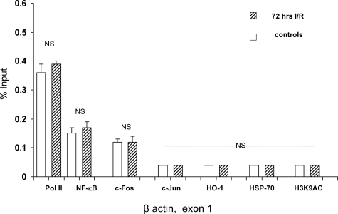
Figure 6.
Assessments performed at a control gene (β-actin, exon 1). I/R did not significantly alter Pol II levels at β-actin, exon 1, serving as a negative control for the data shown at the right of Figure 2. Furthermore, I/R did not impact NF-κB, c-Fos, or c-Jun binding to β-actin, (negative control for the data in Figure 4). Finally, no difference in the extent of trimethylation of histone 3 lysine 4 (H3K4m3) was observed, indicating the relative specificity for data presented in Figure 8. [Note: H3K9AC and H2A.Z variants at β-actin were not assessed.]

