Figure 2.
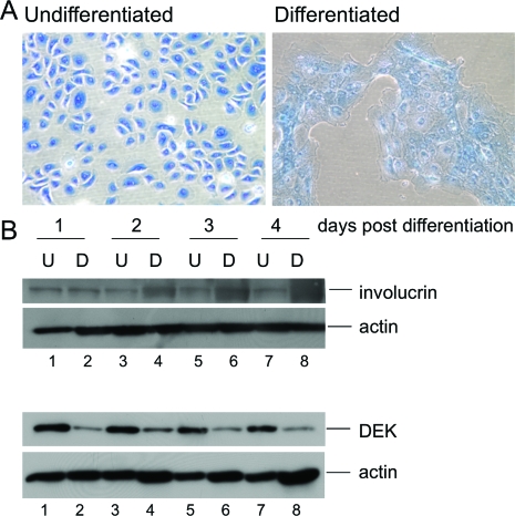
DEK suppression occurs during keratinocyte differentiation in vitro. A: Keratinocyte morphology following calcium addition. Primary human foreskin keratinocytes (HFKs) were either left untreated or subjected to 1 mmol/L CaCl2 and 10% fetal bovine serum to induce differentiation. Cells were fixed and stained with methylene blue. Images were captured 24 hours post treatment. B: Western blot analyses. HFKs were treated as above and total cell protein lysates were harvested between days 1 to 4 post-treatment. Lysates were subjected to Western blot analyses for the detection of DEK, involucrin and actin as a loading control.

