Figure 3.
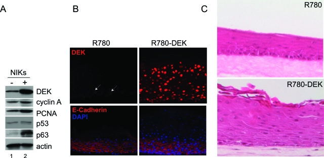
DEK overexpression results in epithelial hyperplasia in organotypic rafts. A: Western blot analyses. NIKs infected with R780 empty vector (−) or DEK (+) were harvested for total cell protein lysates. Equal amounts of protein were subjected to Western blot analyses for the detection of DEK, Cyclin A, p53, p63 and actin. B: Immunofluorescence microscopy. Organotypic raft sections were generated as described in the Materials and Methods, and then incubated with DEK or E-cadherin antibody followed by rhodamine conjugated secondary antibody. Images were captured using an immunofluorescence microscope. C: H&E staining. Sections of NIKs rafts either overexpressing DEK or controls were stained with H&E and images were captured using light microscopy. White arrows denote DEK positive nuclei.

