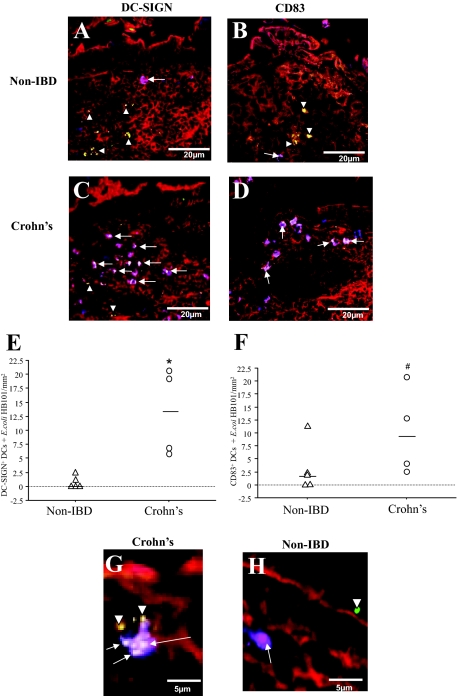
Figure 3.
Enhanced internalization of fluorescent E. coli HB101 by DCs in CD. Sections of distal ileum containing Peyer’s patches from five non-IBD control, and four CD patients were mounted in Ussing chambers. Live GFP E. coli HB101 was added to the mucosal compartment and after 30 minutes of exposure, the tissues were processed for confocal microscopy. Staining of non-IBD control sections illustrated that there were few co-localizations of DC-SIGN+ DCs (A; purple,§ arrows) and CD83+ DCs (B) with E. coli. In CD tissue sections, a higher frequency of both DC-SIGN+ (C) and CD83+ (D) DCs were found to internalize bacteria (white, arrows) in the SED. E. coli was also found either in the extracellular matrix between cells or in cells other than DCs (green, arrowheads). E: Quantification of the co-localized DC-SIGN+ DCs and E. coli HB101 showed significant increase in CD (n = 4) versus non-IBD tissue sections (n = 5). F: Quantification showed that CD83+ DCs co-localization with E. coli were more frequent in CD (n = 4) than in non-IBD (n = 4) tissues. Magnification of the Peyer’s patches showed that in CD (G), the DCs were not only closely associated with the FAE, but were also able to internalize more than one bacterium, in contrast to observations in non-IBD controls (H). From each patient tissue, 10 to 12 sections were examined. Red = F-actin; green = fluorescently-labeled E. coli HB101; §purple = DC-SIGN or CD83 (blue) co-localized with F-actin (red); white = co-localization of DCs with E. coli. *P = 0.01, #P = 0.05.

