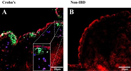
Figure 5.
The FAE cells express and release CCL20. A: Staining for CCL20 (cyan) illustrated that it was expressed and released by the FAE into the SED and lumen of CD tissue sections. Magnification of the SED revealed the recruitment and clustering of DCs in areas where CCL20 release was more prominent (inset). B: In none of the non-IBD controls (stained) did we find FAE expressing CCL20 (representative of three slides from three different patients). Red = F-actin; blue = DC-SIGN; cyan = CCL20. Inset magnification, ×500.

