Figure 1.
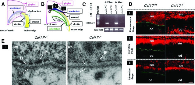
COL17 expression in the tooth of Col17+/+ mice and COL17 absence in the tooth of Col17−/− mice. A, B: Mouse incisors are continuously elongating teeth. In the root of these incisors, ameloblasts (blue) and odontoblasts (green) secrete enamel matrix and dentin, respectively, during the secretory stage (II). I: the pre-secretory stage; II: the secretory stage; III: the maturation stage. C: A RT-PCR assay revealed that Col17 mRNA (488 bp band) was expressed in cultured ameloblasts from Col17+/+ mice (left lane) and Col17+/+ mouse teeth (second right). Col17 mRNA was not expressed in cultured ameloblasts from Col17−/− mice (second left lane) or Col17−/− mouse teeth (right hand lane). D: Immunofluorescence staining for COL17 (green) revealed that COL17 was expressed in the EMJ between ameloblasts and odontoblasts at the pre-secretory stage of a Col17+/+ mouse (upper, left), between ameloblasts and enamel matrix in the secretory stage (middle, left) and in the maturation stage (lower, left) of a Col17+/+ mouse. At the secretory stage, COL17 expression was weak, intermittent, or absent. In Col17−/− mice, no COL17 staining was observed in the EMJ at any stage (right column). am: ameloblast; od: odontblast. Scale bar = 20 μm. E: Ultrastructural features of the basement membrane zone at the pre-secretory stage. Normal hemidesmosomes were seen in the Col17+/+ mouse (left), but hypoplastic, malformed hemidesmosomes were observed in the Col17−/− mice (right). am: ameloblast; LL: lamina lucida; IP: inner attachment plaques; LD :lamina densa. Scale bar = 60 nm.

