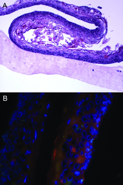
Figure 2.
Expression pattern of L2 and PML in HPV16-transduced organotypic raft cultures. A: H&E stain of a paraffin-embedded HPV16-transduced organotypic raft culture. B: The localization of L2 and PML was examined in a HPV16-transduced organotypic raft culture by co-immunofluorescent staining with RG-1 (green) and PML-specific rabbit antiserum (red). The cellular DNA was stained with DAPI (blue) and the cells imaged by fluorescence microscopy. Original magnifications: ×100 (A); ×400 (B).

