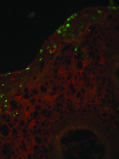
Figure 3.
Expression pattern of HPV6 L2 and PML in condyloma. Immunofluorescent staining of L2 and PML in a frozen section of a florid HPV6-positive condyloma using rabbit antiserum to full-length HPV6 L2 (green) and the monoclonal antibody PG-M3 specific to residues 37 to 51 of human PML (red). The section was imaged by fluorescence microscopy. Original magnification: ×400.

