Figure 1.
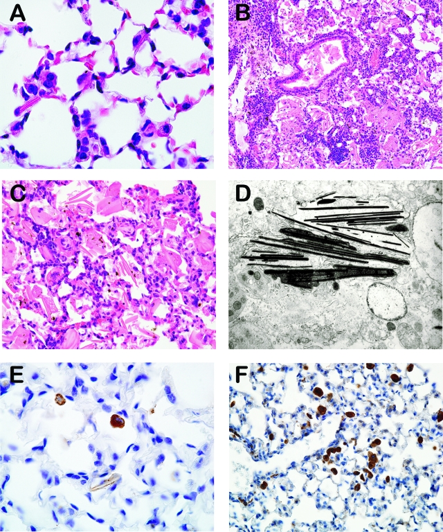
A–F: Histology of macrophage crystalline pneumonia. A: Lung, early crystalline pneumonia, 4-month-old p47phox−/− mouse, H&E, 1000x magnification. B: Lung, severe lesions with abundant crystalline material within alveoli and bronchioles, 14-month-old p47phox−/− mouse, H&E, original magnification ×200. C: Lung, higher magnification demonstrating intra and extracellular giant crystals admixed with macrophages, 14-month-old p47phox−/− mouse, H&E, original magnification ×400. D: Spleen, electron microphotograph of crystalline material showing electron-dense spicules, >12-month-old p47phox−/− mouse (original magnification ×11,880). E: Lung, immunostaining against Ym1/Ym2 protein visualized in pulmonary macrophages with few crystals, 9-week-old p47phox−/− mouse, IHC, original magnification ×1000. F: Lung, immunostaining against macrophage mannose receptor, 9-week-old p47phox−/− mouse, IHC, original magnification ×400.

