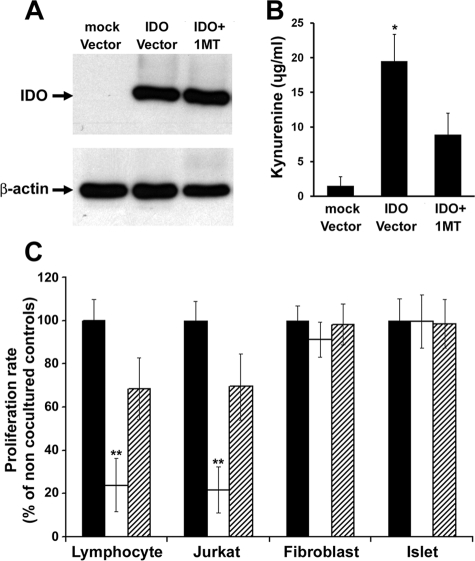
Figure 1.
Selective suppressive effect of IDO on immune versus non immune cells. Mouse fibroblasts were transduced to express IDO using an adenoviral vector. A: IDO protein expression in mock and IDO vector infected cells. The level of kynurenine (the product of IDO mediated tryptophan degradation) was measured in the conditioned media (B). C: Stimulated mouse lymphocytes, Jurkat cells, mouse fibroblasts, and islets were cocultured in two-chamber culture plates with IDO-expressing (open bars) or control fibroblasts (solid bars) for 72 hours. A competitive IDO inhibitor, 1-methyl-tryptophan was added to one set of IDO-expressing cocultures (hatched bars). Cell proliferation rates were measured after 72 hours post-coculture using MTT assay. * denotes significant increase in kynurenine level in IDO vector infected cell conditioned medium compared to the control group (n = 3, P < 0.001); ** denotes significant difference in cell proliferation rate in comparison to the control group (n = 5, P < 0.001).

