Figure 8.
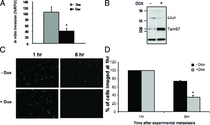
Inhibition of K7M2 in vitro and in vivo metastasis by Tam67. A: In vitro invasion assays of K7M2-Tam67 cells grown in the absence and presence of doxycycline. In vitro invasion was determined by a fluorometric-based cell invasion assay using transwell plates with 8-μm pore size that have been coated with basement membrane matrix. Induction of Tam67 expression with doxycycline significantly inhibited in vitro invasion. B: Western blot analysis showing Tam67 and cJun levels in a pool of K7M2-Tam67 cells prepared for lung colonization assays. Cells were grown in the absence and presence of doxycycline for 48 hours before harvesting for invasion assays. Overexpression of Tam67 results in a decrease in cJun levels as observed in Figure 5B. C: Short term in vivo experimental metastasis imaging of fluorescently labeled K7M2-Tam67 cells grown in the absence and presence of doxycycline for 48 hours before tail-vein injection of the cells into mice. Fluorescent cells located in the lungs of injected animals were photographed at 1 and 6 hours after tail-vein injection. Representative fields from lungs are shown at original magnification ×100. D: Quantification of fluorescence detected in the lungs of eight mice per group injected with control (−dox) and Tam67 expressing (+dox) K7M2 cells. Results shown are the mean ± SEM and are expressed as a percentage of cells in the lungs imaged at 1 hour after tail-vein injection. (*P < 0.05, n = 3).

