Figure 4.
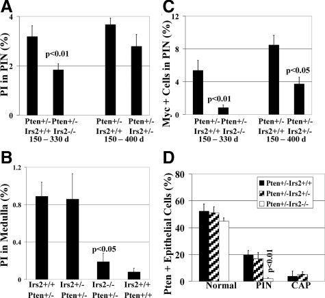
Deletion of Irs2 suppresses epithelial growth, proliferation, and Myc. A: Proliferation index (PI) determined by BrdU immunolabeling of PIN. PI is significantly reduced in Pten+/−Irs2−/− mice (P < 0.01) and trends toward reduction in Pten+/−Irs2+/− mice when compared to the Pten+/−Irs2+/+ group throughout respective study periods. Invasive prostate cancer (not shown) had the highest proliferation index (13.1 ± 1.9%). B: Irs2 deletion suppresses adrenal medulla proliferation attributable to Pten+/−. In adrenal medulla proliferation index is significantly reduced (P < 0.05) in Pten+/−Irs2−/− but not in Pten+/−Irs2+/− mice versus Pten+/−Irs2+/+ controls. C: Nuclear Myc immunolabeling is significantly reduced in Pten+/−Irs2−/− (P < 0.01) and Pten+/−Irs2+/− (P < 0.05) mice compared Pten+/−Irs2+/+ controls. Invasive prostate cancer (9.38 ± 1.97%) has the highest number of Myc-positive cells (data not shown). D: Pten expression is markedly reduced in Pten+/−Irs2−/− PIN lesions. Comparison of percentage of Pten-positive prostatic epithelial cells in normal tissue, PIN, and CAP of Pten+/−Irs2+/+, Pten+/−Irs2+/−, and Pten+/−Irs2−/− mice. Notably, PIN of Pten+/−Irs2−/− mice shows nearly complete Pten loss of expression with on average 2.1 ± 0.6% cells immunoreactive for Pten, which is a significant reduction compared to PIN in Pten+/−Irs2+/+ controls (P < 0.01).

