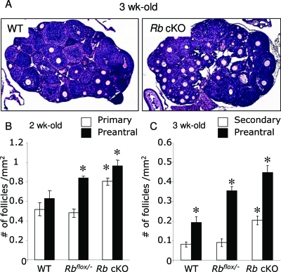
Figure 2.
Histological Analysis and Follicle Counts of the Ovaries of Prepubertal Control and Rb cKO Mice
A, Ovaries from 3-wk-old WT and Rbflox/−Amhr2Cre+ (Rb cKO) mice. Note the increased amount of growing follicles present in the Rb cKO ovary. B and C, Follicle counts of 2-wk-old (B) and 3-wk-old (C) WT and Rb cKO mice. Follicles were counted in five histological sections derived from four to five ovarian samples and the number of follicles per square millimeter was calculated as described in Materials and Methods. Two-week-old Rb cKO ovaries show a significantly higher number of primary and preantral (multilayer) follicles compared with WT ovaries, whereas 3-wk-old Rb cKO ovaries show a significantly higher number of secondary and preantral follicles compared with WT. Only follicular stages that displayed significant differences are shown. For counts in all follicle stages, please refer to supplemental Fig. 1, A and B. Magnification, ×50. *, P < 0.05.

