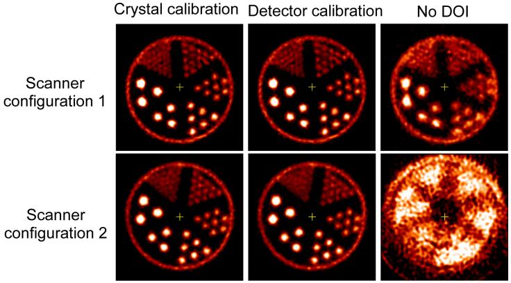
Figure 8.
Images of a hot rod phantom reconstructed with (left) a linear crystal DOI calibration and (middle) a linear detector DOI calibration, and (right) without DOI information. The rod diameters are 0.75, 1.0, 1.35, 1.70, 2.0 and 2.4 mm. Rod to rod separation is twice the rod diameter. The images were reconstructed by FBP with a Shepp-Logan filter cut off at 0.5 of the Nyquist frequency.

