Figure 1.
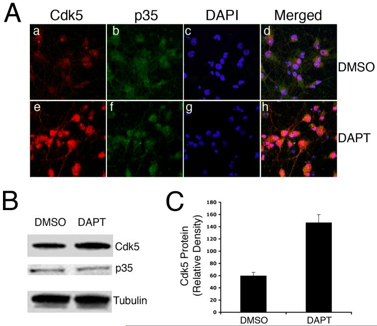
DAPT induces upreguation of cdk5. E18 rat embryonic cortical neurons were cultured for 7 d in B27/neurobasal medium and then treated with either DMSO or 10 μM DAPT for 24h. Cells were lysed for immunoblotting or fixed for immunocytochemistry (ICC). (A) Fixed cells were immunostained for cdk5 (a, e) and p35 (b, f). Nuclei are stained with DAPI (c, g). Immunostaining for cdk5, p35 and DAPI are merged (d, h). (B) A representative immunoblot shows cdk5, p35 levels in the DMSO- and DAPT-treated neurons. Alpha-tubulin levels are shown as loading controls. (C) Quantitation of cdk5 expression in the control, DMSO- treated and DAPT-treated cells. Data are derived from three separate experiments.

