Figure 2.
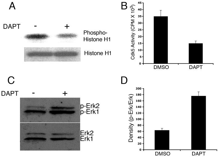
DAPT downregulates cdk5 activity and activates MAPK (Erk1/2). (A) Cdk5 kinase activity is shown for DMSO- and DAPT-treated neuronal extracts in a representative autoradiogram. Upper panel shows the autoradiogram of phosphorylated Histone H1 by the immunoprecipitated cdk5 from the lysates. Lower panel shows Coomassie blue staining of the Histone H1 substrate used for the kinase assay. (B) Densitometric analyses of phosphorylated Histone H1 from three different autoradiograms show the difference in cdk5 activity between DMSO- and DAPT-treated neurons. (C) A representative immunoblot shows p-Erk (upper panel) and total Erk levels in cortical neurons treated with DMSO- and DAPT.

