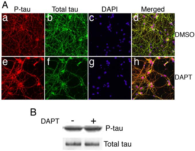
Figure 3.
DAPT affects distribution of tau protein. E18 rat embryonic cortical neurons were cultured for 7 d in B27/neurobasal medium and then treated with either DMSO (a-d) or 10 μM DAPT (e-h) for 24 h. Cells were lysed for immunoblotting or fixed for immunocytochemistry (ICC). (A) Fixed cells were immunostained for phospho-tau (p-tau) (a, e) and total tau (b, f). Nuclei are stained with DAPI (c-g). Merged image of p-tau, total tau and DAPI staining of DMSO-treated cells is shown (d) and that DAPT-treated cells (h) are shown. (B) Immunoblot shows p-tau and total tau levels in the DMSO- and DAPT-treated neurons.

