Figure 2.
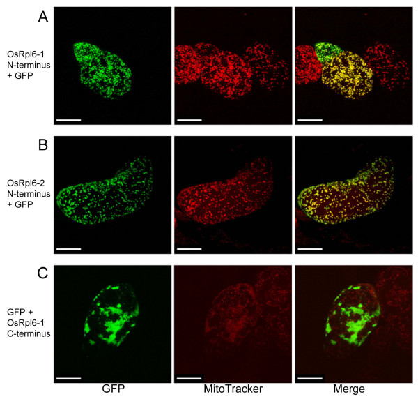
Subcellular localization of GFP fusion proteins in tobacco BY-2 cells. Constructs were fused to GFP cDNA at the following positions: (A) the N-terminal coding region of OsRpl6-1 was fused to 5'-upstream position of GFP, (B) the N-terminal coding region of OsRpl6-2 was fused to 5'-upstream position of GFP, and (C) the C-terminal coding region of OsRpl6-1 was fused to 3'-downstream position of GFP. Representative images are shown. Left: GFP fluorescence. Center: fluorescence of a mitochondrial-specific dye, MitoTracker Red. Right: merging of both signals. Scale bar = 20 μm. In the right panel of Figure 2A, some of the GFP fluorescence within a small cell behind the central cell did not co-localize with mitochondria. The GFP fluorescence from the behind cell would have been overexposed, probably because its GFP expression had been much more enhanced than that in the central cell.

