
Figure 2. VSP citrullination is likely mediated by the PAD activity of gADI.
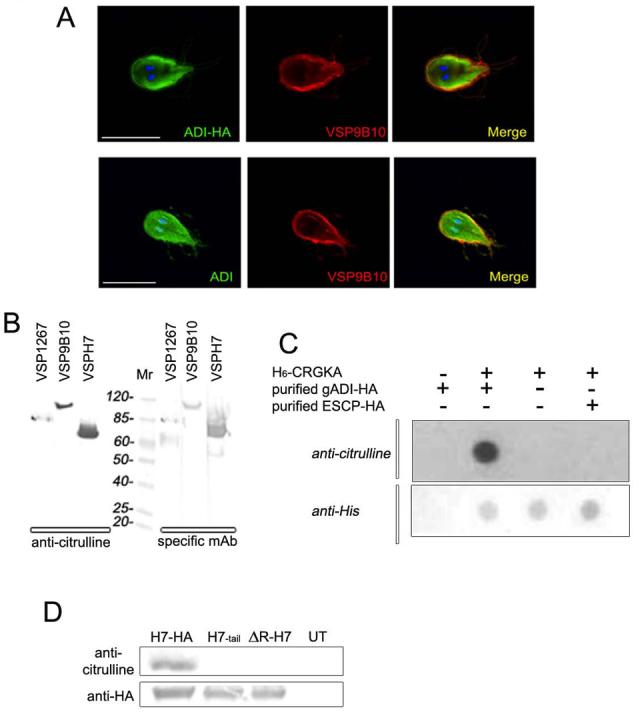
(A) Confocal direct IFA was performed on permeabilized cells, showing a cytoplasmic distribution of gADI-HA (green) by using FITC-labeled anti-HA mAb, and its partial colocalization (yellow in merge) with VSP9B10 (red) underneath the plasma membrane of the transgenic trophozoite (top panels). The same result was obtained by using Alexa 488-anti-gADI in wild-type cells (bottom panels). Texas Red-9B10 mAb was used to visualize VSP9B10. Nuclei are stained with DAPI (blue). Scale bar represents 10 μm.
(B) Western blotting of Giardia homogenates expressing different VSPs reacted with anti-citrulline pAb (left pannel). The same filter membranes were stripped, cut, and reacted with 5C1, 9B10, and G10/4 mAbs against VSP1267, VSP9B10, and VSPH7, respectively (right panel), indicating that these VSPs are citrullinated. Mr is molecular mass expressed in kDa.
(C) Dot-blotting to detect citrullination of the H6-CRGKA peptide after incubation with the purified recombinant ADI-HA. A non-related purified enzyme ESCP-HA was used as a negative control. Dot-blotting to detect H6-CRGKA was performed by using anti-H6mAb.
(D) Specific citrullination of the CRGKA tail is shown (top panel). Western blotting using anti-citrulline pAb performed after immunoprecipitation with anti-HA mAb of H7-transgenic trophozoites. The presence of H7-HA, H7-tail, and ΔR-H7 after immunoprecipitation was analyzed by using anti-HA mAb labeled with alkaline phosphatase (bottom panel).