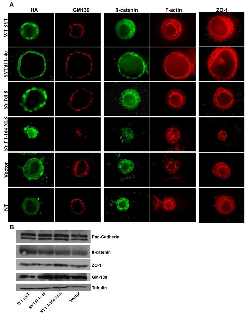
Fig. 2. Integrity of epithelial polarity in SYT expressants.
Confocal microscopy of MDCK cells infected with WT SYT, the SYT deletion (dl) mutants, and grown in collagen for 7 days. (A) Cysts were double stained with antibodies to the apical Golgi protein GM130 (red) and with anti-HA antibody (to visualize the expressed SYT cDNAs). Polarization was also assessed by the luminal localization of an actin ring (red) at the apical surface, the basolateral marker β-catenin (green) and the apical marker ZO-1 (red). (B) Cellular levels of polarity markers in the monolayers of SYT dl mutants-expressing MDCK cells.

