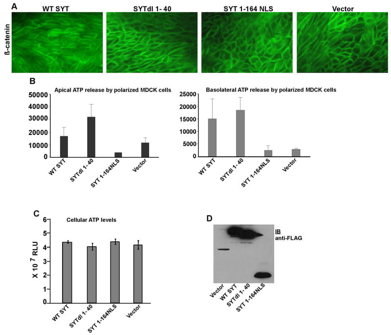
Fig. 4. Effect of SYT mutants on apical and basolateral ATP release by polarized MDCK cells.
(A) Immunostaining of β-catenin in polarized MDCK cells expressing WT SYT and SYT dl mutants. (B) ATP released from apical and basolateral surfaces of MDCK cells expressing WT SYT and various SYTdl mutants. The values were recorded as Relative Light Units (RLU). (C) Steady state levels of intracellular ATP produced in control vector-, WT SYT-, SYTdl 1–40-, and SYT 1–164NLS-polarized MDCK cells. Measurements were reproduced in 3 independent experiments (n=3), each conducted in triplicates. (D) Expression levels of WT SYT, SYTdl1–40, and SYT1-164NLS in polarized MDCK cells visualized with the anti-FLAG antibody. The protein band in the pOZ vector-lane is the irrelevant vector-generated polypeptide that disappears upon subcloning of SYT cDNAs.

